By Ihab R. Kamel, Elmar M. Merkle
ISBN-10: 0521194865
ISBN-13: 9780521194860
Physique MR Imaging at 3.0 Tesla is a pragmatic textual content permitting radiologists to maximize the advantages of excessive box 3T MR platforms in more than a few physique functions. It explains the actual rules of MR imaging utilizing 3T magnets, and the variations among 1.5T and 3T whilst utilized extracranially. The book's organ-based process makes a speciality of optimized innovations, supplying urged protocols for the most owners of 3T MRI platforms. All significant thoracic and belly organs are lined, together with breast, center, liver, pancreas, the GI tract, kidneys, prostate and feminine pelvic organs. stomach and pelvic MR angiography and MRCP also are mentioned. Protocol optimization, visual appeal of artifacts and novel functions utilizing 3T are emphasised. Written and edited by way of specialists within the box, physique MR Imaging at 3.0 Tesla courses radiologists in optimizing imaging protocols for 3T MR structures, decreasing artifacts and making a choice on some great benefits of utilizing 3T in physique applications
''Body MR Imaging at 3.0 Tesla is a realistic textual content permitting radiologists to maximize some great benefits of excessive box 3T MR structures in a variety of physique purposes. It explains the actual ideas of MR imaging utilizing 3T magnets, and the variations among 1.5T and 3T whilst utilized extracranially. The book's organ-based method specializes in optimized recommendations, delivering advised protocols for the most owners of 3T MRI structures. All significant thoracic and stomach organs are coated, together with breast, center, liver, pancreas, the GI tract, kidneys, prostate and feminine pelvic organs. belly and pelvic MR angiography and MRCP also are mentioned. Protocol optimization, visual appeal of artifacts and novel functions utilizing 3T are emphasised. Written and edited through specialists within the box, physique MR Imaging at 3.0 Tesla publications radiologists in optimizing imaging protocols for 3T MR platforms, decreasing artifacts and making a choice on the benefits of utilizing 3T in physique applications''--Provided by way of writer. Read more... physique MRI at 3T: uncomplicated issues approximately artifacts and defense / Kevin J. Chang and Ihab R. Kamel -- Novel acquisition concepts which are facilitated via 3T / Hiroumi D. Kitajima, Puneet Sharma, Daniel R. Kayolyi and Diego R. Martin -- Breast MR imaging / Savannah C. Partridge, Habib Rahbar and Constance D. Lehman -- Cardiac MR imaging / Christopher J. Francois, Oliver Wieben and Scott B. Reeder -- stomach and pelvic MR angiography / Henrik J. Michaely -- Liver MR imaging at 3T: demanding situations and possibilities / Elizabeth M. Hecht and Bachir Taouli -- MR imaging of the pancreas / Sang Soo Shin, Chang Hee Lee, Rafael O. P. de Campos and Richard C. Semelka -- MR imaging of the adrenal glands / Daniele Marin and Elmar M. Merkle -- Magnetic resonance cholangiopancreatography / Byung Ihn Choi and Jeong Min Lee -- MR imaging of small and massive bowel / M. L. W. Zeich, M. P. van der Paardt, A. J. Nederveen and J. Stoker -- MR imaging of the rectum, 3T vs 1.5T / Monique Maas, Doenja M. J. Lambregts and Regina G. H. Beets-Tan -- Imaging of the kidneys and MR urography at 3T / John R. Leyendecker -- MR imaging and MR-guided biopsy of the prostate at 3T / Katarzyna J. Macura and Jurgen J. Futterer -- woman pelvic imaging at 3T / Darcy J. Wolfman and Susan M. Ascher
Read or Download Body MR imaging at 3 Tesla PDF
Best imaging systems books
Investigations of Field Dynamics in Laser Plasmas with Proton Imaging
Laser-driven proton beams are nonetheless of their infancy yet have already got a few notable attributes in comparison to these produced in traditional accelerators. One such characteristic is the ordinarily low beam emittance. this enables very good solution in imaging functions like proton radiography. This thesis describes a unique imaging method - the proton streak digital camera - that the writer constructed and primary used to degree either the spatial and temporal evolution of ultra-strong electric fields in laser-driven plasmas.
Mathematical morphology in image processing
Education structuring parts in morphological networks / Stephen S. Wilson -- effective layout suggestions for the optimum binary electronic morphological filter out: percentages, constraints, and structuring-element libraries / Edward R. Dougherty and Robert P. Loce -- Statistical homes of discrete morphological filters / Jaakko Astola, Lasse Koskinen, and Yrjö Neuvo -- Morphological research of pavement floor / Chakravarthy Bhagvati, Dimitri A.
The overseas Acoustical Imaging Symposium has been held regularly considering the fact that 1968 as a special discussion board for complicated study, selling the sharing of know-how, advancements, tools and concept between all parts of acoustics. The interdisciplinary nature of the Symposium and the vast overseas participation are of its major strengths.
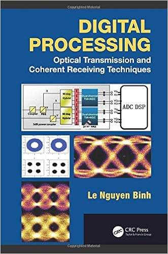
Digital Processing: Optical Transmission and Coherent Receiving Techniques
With coherent blending within the optical area and processing within the electronic area, complicated receiving suggestions using ultra-high pace sampling premiums have stepped forward greatly over the past few years. those advances have introduced coherent reception structures for lightwave-carried info to the following degree, leading to ultra-high capability worldwide internetworking.
- Colour Image Processing Handboook
- Digital and Analog Fiber Optic Communications for CATV and FTTx Applications (SPIE Press Monograph Vol. PM174) (Press Monograph)
Extra resources for Body MR imaging at 3 Tesla
Sample text
Matsuoka A, Minato M, Harada M, et al. 5-tesla diffusion-weighted imaging in the visibility of breast cancer. Radiat Med 2008; 26: 15–20. 15. Lo GG, Ai V, Chan JK et al. Diffusion-weighted magnetic resonance imaging of breast lesions: first experiences at 3T. Comput Assist Tomogr 2009; 33(1): 63–9. 16. Bolan PJ, Nelson MT, Yee D, Garwood M. Imaging in breast cancer: magnetic resonance spectroscopy. Breast Cancer Res 2005; 7: 149–52. 17. Sinha S, Sinha U. Recent advances in breast MRI and MRS. NMR Biomed 2009; 22: 3–16.
This chapter will review technical considerations for performing CMR at 3T, including the impact of 3T on SNR, relaxation times, chemical shift and susceptibility, specific absorption rate (SAR), dielectric effects (magnetic field inhomogeneity), bSSFP imaging, and parallel imaging. This is followed by a discussion of specific CMR sequences and their performance at 3T. Technical considerations for CMR at 3T Signal-to-noise ratio (SNR) The increase in SNR is one of the most appealing reasons for imaging at a higher field strength.
Detection, but continues to experience moderate specificity, ranging from 37% to 88%. 5T have primarily focused on improved differentiation of benign lesions from malignant lesions. As the current clinical breast MR imaging approach is to assess lesion morphology and lesion kinetics for diagnostic classification, initial research has centered on potential improvements that higher field strength may provide for these two parameters. Lesion identification is primarily dependent on the contrast-to-noise ratio (CNR), which is dependent upon relaxation times of breast tissue and gadoliniumbased contrast agents used for breast MRI.


