By Devin Binder, D. Sonne, Nancy Fischbein
ISBN-10: 1588904024
ISBN-13: 9781588904027
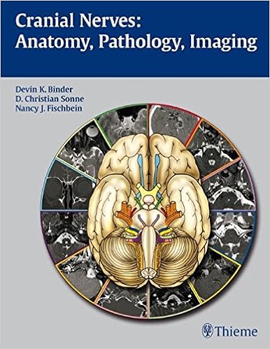
This fantastically illustrated e-book combines a close exposition of the anatomy and serve as of the cranial nerves with sensible insurance of scientific recommendations for the evaluate and differential prognosis of cranial nerve disorder. An introductory bankruptcy presents a short evaluate of cranial nerve anatomy and serve as, cranium base anatomy, category of pathologies, and imaging methods. all of the twelve chapters that stick with is dedicated to in-depth assurance of a special cranial nerve. those chapters open with exact dialogue of many of the capabilities of every nerve and general anatomy. The authors then describe universal lesions and current a chain of situations which are complemented by way of CT pictures and MRIs to demonstrate sickness entities that lead to cranial nerve dysfunction.Highlights:-Concise descriptions in a bulleted define layout let swift analyzing and review-Tables synthesize key details concerning anatomy, functionality, pathology, and imaging-More than three hundred top quality illustrations and state of the art CT and MR photographs exhibit very important anatomic recommendations and pathologic findings-Pearls emphasize scientific info and key imaging findings for analysis and treatment-Appendices comprise certain details on brainstem anatomy, scholar and eye circulation keep an eye on, parasympathetic ganglia, and cranial nerve reflexesThis ebook is an imperative reference for training physicians and trainees in neurosurgery, neurology, neuroradiology, radiology, and otolaryngology-head and neck surgical procedure. it's going to additionally function a priceless source for college kids trying to achieve a great realizing of the anatomy, functionality, and pathology of the cranial nerves.
Read or Download Cranial Nerves: Anatomy, Pathology, Imaging PDF
Best neurosurgery books
Musculoskeletal Diseases: Diagnostic Imaging and Interventional Techniques
This e-book represents a condensed model of the 20 issues facing imaging prognosis and interventional treatments in musculoskeletal ailments. The disease-oriented issues surround all of the proper imaging modalities together with X-rays expertise, nuclear drugs, ultrasound and magnetic resonance, in addition to image-guided interventional innovations.

Erythropoietin and the Nervous System
Erythropoietin (EPO) is a chemokine hormone that's commonly allotted during the physique. as well as its conventional position as a hormone that stimulates crimson blood cellphone construction, in recent times many laboratories have proven that EPO can act as a neuroprotective compound in various damage paradigms within the worried process.
Percutaneous Vertebroplasty is a concise and up to date reference that info the necessities for constructing a latest medical lab, settling on sufferers, adequately acting the method and fending off pitfalls which are in general encountered. Over ninety five photos, specifically created for this publication, give you the reader with distinctive examples of the way each one element of the technique is played in an comprehensible step-by-step structure.
Electroceuticals: Advances in Electrostimulation Therapies
This e-book covers contemporary advances within the use of electrostimulation cures in flow issues, epilepsy, inflammatory bowel ailment, reminiscence and cognition, problems of cognizance, foot drop, dysphagia, mind harm, headache, center failure, listening to loss, and rheumatoid arthritis. It describes concepts similar to vagus nerve stimulation, deep mind stimulation, and electric stimulation of the pharyngeal nerve.
- Psychosurgery: New Techniques for Brain Disorders
- Neuroscience PreTest Self-Assessment and Review, Sixth Edition (PreTest Basic Science)
- Seizures in critical care : a guide to diagnosis and therapeutics
- Balloon Kyphoplasty
- Neuro-Oncology of CNS Tumors
Additional info for Cranial Nerves: Anatomy, Pathology, Imaging
Example text
Oculomotor palsy, gaze paralysis, and ipsilateral ataxia. 3 Oculomotor Nerve Lesions In the Subarachnoid Space (asternal Segment of Nerve) • Ischemia. Isolated oculomotor palsy with pupillary spar- • • • • • • • • ing is usually indicative of ischemic oculomotor palsy (caused by diabetes, hypertension, atherosclerosis). Diabetic third nerve palsy is usually painfu~ develops over a few hours, and has a good prognosis for recovery. Imaging is not indicated in the clinical setting of a classic diabetic oculomotor palsy.
5} is 1otated below the supradli- asmmc ~cess of the third veotride, lamina terminalis. • Vastular relationships: Posterior oommunkatlng artery (PCommA) located Inferior to optic tracts In suprasellar posterior cerebral artery (PCA) and basal vein Rosenthal apposed to optit tracts in perimesentephalic astern. or cistern and anterior coll'ID'Ils:sure; tbe pituitary stalk Is ~ alely posterior. Retinal Targets • There are four brain regions that directly receive visual Information from the retina (1'13.
Correction should not be undatalcen until rec:overy Is cmnplete; In most cases the defidt sUblllzes within -6 montbs. ology. More common than all other lntncranial tumon wmblned Occur In 10 to 30l of all patients with systemic canur. • Cllnlad~ Prefentwlthheadadle, back palo. lOcal neuroiosk deficit, and{or seizures deperuliog on location. Rf. 15 Axial post1joid*'lum n-welghtld lmagl! nsely enhindng mus centered on the left superior c:ollltulus (curow). Mild perlphenl ·l~lty Is campatible with adjK2nt vasogenk: edema.