By Klaus Dieter Maria Resch
ISBN-10: 3540303197
ISBN-13: 9783540303190
ISBN-10: 3540425055
ISBN-13: 9783540425052
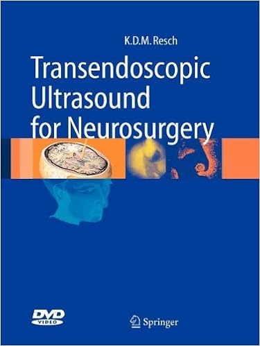
Endoscopic neurosurgery has no longer but reached the security and applicability of microsurgery. Endo-neuro-sonography is a brand new process aimed toward making endoscopy more secure by means of real-time imaging and navigation capability (brain-radar). The endo-neurosonographic snapshot is a sonographic test on the tip of the endoscope (mini-CT) providing additional info to the endoscopic view. Endo-Neuro-Sonography is the 1st e-book in this new subject, featuring ideas and kit, anatomical positive factors, and the 1st medical sequence. Schematic drawings, tables and 237 figures (122 in colour) supply an exact review of this system. the ultimate bankruptcy is facing destiny techniques on minimally invasive neurosurgery and particular connection with ergonomics in neurosurgery.
Read or Download Transendoscopic Ultrasound for Neurosurgery PDF
Similar neurosurgery books

Musculoskeletal Diseases: Diagnostic Imaging and Interventional Techniques
This booklet represents a condensed model of the 20 themes facing imaging analysis and interventional treatments in musculoskeletal illnesses. The disease-oriented subject matters surround all of the appropriate imaging modalities together with X-rays expertise, nuclear medication, ultrasound and magnetic resonance, in addition to image-guided interventional suggestions.

Erythropoietin and the Nervous System
Erythropoietin (EPO) is a chemokine hormone that's extensively allotted through the physique. as well as its conventional function as a hormone that stimulates pink blood mobilephone creation, lately many laboratories have proven that EPO can act as a neuroprotective compound in various harm paradigms within the anxious procedure.
Percutaneous Vertebroplasty is a concise and updated reference that info the necessities for constructing a latest scientific lab, settling on sufferers, accurately appearing the technique and warding off pitfalls which are as a rule encountered. Over ninety five pictures, in particular created for this ebook, give you the reader with specific examples of ways every one element of the strategy is played in an comprehensible step-by-step structure.
Electroceuticals: Advances in Electrostimulation Therapies
This booklet covers contemporary advances within the use of electrostimulation remedies in flow issues, epilepsy, inflammatory bowel disorder, reminiscence and cognition, issues of attention, foot drop, dysphagia, mind damage, headache, middle failure, listening to loss, and rheumatoid arthritis. It describes recommendations reminiscent of vagus nerve stimulation, deep mind stimulation, and electric stimulation of the pharyngeal nerve.
- Management of Neurological Disorders
- Management of Neurological Disorders
- The Cranial Nerves: Anatomy Imaging Vascularisation
- Neuro-Oncology: The Essentials
- Complications in Head and Neck Surgery with CD Image Bank, 2e
Extra resources for Transendoscopic Ultrasound for Neurosurgery
Sample text
105 Indications . . . . . . . . . . . . . . . . . . . . 106 Conclusions . . . . . . . . . . . . . . . . . . . 109 Intraoperative endoneurosonographic (ENS) imagings prepared during surgery on 52 selected patients between April 1996 and July 2000 were examined. There were 23 female and 29 male patients, and their mean age was 42 (2–69) years. In most cases Aloka (Aloka Deutschland, Düsseldorf, Germany) sono equipment was used because equipment supplied by this company had yielded superior imaging results in Objective After transendoscopic sono-catheters had been tested in the laboratory for imaging characteristics and practicability (chap.
The sono-probe touches the pons (6). In the clivus (2) a small sphenoid sinus (10) becomes visible the fourth ventricle (2). Ventrally the surface of the rhombencephalon with facial colliculus (4) is visible, while dorsally the choroid plexus (3) is present Fig. 58. Cisterna magna level. The sono-probe (1) is placed at the cisterna magna (5) and has made contact with the dorsal surface of the medulla (2). Ventral to the medulla (2), the premedullary cistern (3) and the clivus (4) are visible. Dorsally, the tonsils (6) and lobus biventer (7) are present Fig.
The thalamostriate vein (5) is crossing the lamina affixa (6). The tip of the probe (1) can be accurately controlled by the endoscope and can be pushed ahead into the working channel of the endoscope, independently visualizing parts that are invisible to the endoscope of Monro (2) formed by the fornix (4) and the choroid plexus (3). The scan represents the pellucid septum (5) Fig. 7. The sono-probe (1) is pushed into the foramen of Monro (2) formed by the fornix (3) touching the intermediate mass (4).