By Gustaaf van Tendeloo, Dirk van Dyck, Stephen J. Pennycook
ISBN-10: 3527317066
ISBN-13: 9783527317066
ISBN-10: 3527641866
ISBN-13: 9783527641864
Content material:
Chapter 1 Transmission Electron Microscopy (pages 9–44): Marc De Graef
Chapter 2 Atomic answer Electron Microscopy (pages 45–79): Prof. Dirk Van Dyck
Chapter three Ultrahigh?Resolution Transmission Electron Microscopy at adverse round Aberration (pages 81–107): Knut W. city, Juri Barthel, Lothar Houben, Chun?Lin Jia, Markus Lentzen, Andreas Thust and Karsten Tillmann
Chapter four Z?Contrast Imaging (pages 109–152): Prof. Dr. Stephen J. Pennycook, Anrew R. Lupini, Albina Y. Borisevich and Mark P. Oxley
Chapter five Electron Holography (pages 153–220): Hannes Lichte
Chapter 6 Lorentz Microscopy and Electron Holography of Magnetic fabrics (pages 221–251): Rafal E. Dunin?Borkowski, Takeshi Kasama, Marco Beleggia and Giulio Pozzi
Chapter 7 Electron Tomography (pages 253–279): Paul Anthony Midgley and Sara Bals
Chapter eight Statistical Parameter Estimation thought – a device for Quantitative Electron Microscopy (pages 281–308): Sandra Van Aert
Chapter nine Dynamic Transmission Electron Microscopy (pages 309–343): Nigel D. Browning, Geoffrey H. Campbell, James E. Evans, Thomas B. LaGrange, Katherine L. Jungjohann, Judy S. Kim, Daniel J. Masiel and Bryan W. Reed
Chapter 10 Transmission Electron Microscopy as Nanolab (pages 345–374): Frans D. Tichelaar, Marijn A. van Huis and Henny W. Zandbergen
Chapter eleven Atomic?Resolution Environmental Transmission Electron Microscopy (pages 375–403): Pratibha L. Gai and Edward D. Boyes
Chapter 12 Speckles in pictures and Diffraction styles (pages 405–435): Michael M. J. Treacy
Chapter thirteen Coherent Electron Diffractive Imaging (pages 437–472): J. M. Zuo and Weijie Huang
Chapter 14 pattern coaching suggestions for Transmission Electron Microscopy (pages 473–498): Vasfi Burak Ozdol, Vesna Srot and Peter A. van Aken
Chapter 15 Scanning Probe Microscopy – background, heritage, and state-of-the-art (pages 499–538): Ralf Heiderhoff and Ludwig Josef Balk
Chapter sixteen Scanning Probe Microscopy – Forces and Currents within the Nanoscale global (pages 539–614): Brian J. Rodriguez, Roger Proksch, Peter Maksymovych and Sergei V. Kalinin
Chapter 17 Scanning Beam tools (pages 615–643): David Joy
Chapter 18 basics of the concentrated Ion Beam process (pages 645–671): Nan Yao
Chapter 19 Low?Energy Electron Microscopy (pages 673–696): Ernst Bauer
Chapter 20 Spin?Polarized Low?Energy Electron Microscopy (pages 697–707): Ernst Bauer
Chapter 21 Imaging Secondary Ion Mass Spectroscopy (pages 709–744): Katie L. Moore, Markus Schroder and Chris R. M. Grovenor
Chapter 22 smooth X?Ray Imaging and Spectromicroscopy (pages 745–791): Adam P. Hitchcock
Chapter 23 Atom Probe Tomography: precept and purposes (pages 793–832): Frederic Danoix and Francois Vurpillot
Chapter 24 sign and Noise greatest probability Estimation in MRI (pages 833–853): Jan Sijbers
Chapter 25 3?D floor Reconstruction from Stereo Scanning Electron Microscopy photos (pages 855–876): Shafik Huq, Andreas Koschan and Mongi Abidi
Chapter 26 Nanoparticles (pages 877–960): Miguel Lopez?Haro, Juan Jose Delgado, Juan Carlos Hernandez?Garrido, Juan de Dios Lopez?Castro, Cesar Mira, Susana Trasobares, Ana Belen Hungria, Jose Antonio Perez?Omil and Jose Juan Calvino
Chapter 27 Nanowires and Nanotubes (pages 961–993): Yong Ding and Zhong Lin Wang
Chapter 28 Carbon Nanoforms (pages 995–1070): Carla Bittencourt and Prof. Gustaaf Van Tendeloo
Chapter 29 Metals and Alloys (pages 1071–1097): Dominique Schryvers
Chapter 30 In situ Transmission Electron Microscopy on Metals (pages 1099–1151): J. Th. M. De Hosson
Chapter 31 Semiconductors and Semiconducting units (pages 1153–1178): Hugo Bender
Chapter 32 complicated Oxide fabrics (pages 1179–1212): Maria Varela, Timothy J. Pennycook, Jaume Gazquez, Albina Y. Borisevich, Sokrates T. Pantelides and Prof. Dr. Stephen J. Pennycook
Chapter 33 software of Transmission Electron Microscopy within the study of Inorganic Photovoltaic fabrics (pages 1213–1246): Yanfa Yan
Chapter 34 Polymers (pages 1247–1272): Joachim Loos
Chapter 35 Ferroic and Multiferroic fabrics (pages 1273–1301): Ekhard Salje
Chapter 36 Three?Dimensional Imaging of Biomaterials with Electron Tomography (pages 1303–1333): Montserrat Barcena, Roman I. Koning and Abraham J. Koster
Chapter 37 Small natural Molecules and better Homologs (pages 1335–1380): Ute Kolb and Tatiana E. Gorelik
Read or Download Handbook of Nanoscopy, Volume 1&2 PDF
Best imaging systems books
Investigations of Field Dynamics in Laser Plasmas with Proton Imaging
Laser-driven proton beams are nonetheless of their infancy yet have already got a few awesome attributes in comparison to these produced in traditional accelerators. One such characteristic is the commonly low beam emittance. this enables first-class solution in imaging purposes like proton radiography. This thesis describes a unique imaging method - the proton streak digicam - that the writer built and primary used to degree either the spatial and temporal evolution of ultra-strong electric fields in laser-driven plasmas.
Mathematical morphology in image processing
Education structuring components in morphological networks / Stephen S. Wilson -- effective layout recommendations for the optimum binary electronic morphological clear out: chances, constraints, and structuring-element libraries / Edward R. Dougherty and Robert P. Loce -- Statistical homes of discrete morphological filters / Jaakko Astola, Lasse Koskinen, and Yrjö Neuvo -- Morphological research of pavement floor situation / Chakravarthy Bhagvati, Dimitri A.
The foreign Acoustical Imaging Symposium has been held regularly due to the fact 1968 as a distinct discussion board for complicated learn, selling the sharing of know-how, advancements, equipment and thought between all components of acoustics. The interdisciplinary nature of the Symposium and the broad foreign participation are of its major strengths.
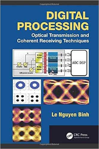
Digital Processing: Optical Transmission and Coherent Receiving Techniques
With coherent blending within the optical area and processing within the electronic area, complex receiving ideas utilising ultra-high pace sampling charges have stepped forward drastically during the last few years. those advances have introduced coherent reception platforms for lightwave-carried info to the subsequent degree, leading to ultra-high capability worldwide internetworking.
- Smart CMOS Image Sensors and Applications (Optical Science and Engineering)
- Computer design of diffractive optics
- Fiber Bragg Gratings (Optics and Photonics)
- Digital Signal Processing (2007)
- Handbook of MRI Pulse Sequences
- PET-TC nella pratica clinica
Extra resources for Handbook of Nanoscopy, Volume 1&2
Sample text
27. 28. 29. 30. 31. tomography, Handbook of Nanoscopy, Wiley-VCH Verlag GmbH, Weinheim. P. (1994) Electron source brightness and degeneracy from Fresnel fringes in field emission point protection microscopy. J. Vac. Sci. Technol. A, 12, 543–547. D. et al. (2012) Dynamic transmission electron microscopy, Handbook of Nanoscopy, Wiley-VCH Verlag GmbH, Weinheim. Yao, N. (2012) Fundamentals of the focused ion beam system, Handbook of Nanoscopy, Wiley-VCH Verlag GmbH, Weinheim. H. M. (2010) 4D Electron Microscopy Imaging in Space and Time, Imperial College Press, London.
Iijima being one of the pioneers in the field of solid state chemistry with his high-resolution studies of Nb2 O5 -based materials [8]. Independently, atomic resolution images of heavy atoms on a carbon support were obtained by Crewe and coworkers in Chicago using a high-resolution scanning transmission electron microscope (STEM) [9]. 1 nm. At this value, the limit seemed to be reached since spherical aberration and chromatic aberration (and often also the sample) limited further progress. However, the introduction of spherical-aberration-corrected lenses [10] opened a new world of subangstrom resolution and improved signal-to-noise ratio.
2012) Statistical parameter estimation theory – a tool for quantitative electron microscopy, Handbook of Nanoscopy, Wiley-VCH Verlag GmbH, Weinheim. Rose, H. (1990) Outline of a spherically corrected semiplanatic medium-voltage transmission electron microscope. Optik, 85, 19–24. Van Dyck, D. (2012) Atomic resolution electron microscopy, Handbook of Nanoscopy, Wiley-VCH Verlag GmbH, Weinheim. Pennycook, S. (2012) Z contrast imaging, Handbook of Nanoscopy, Wiley-VCH Verlag GmbH, Weinheim. Botton, G.